
Description

Price: ₹742.00
(as of Jul 19, 2025 04:52:11 UTC – Details)

This book has been written in a concise and easily assimilable style to enable rapid understanding of the mechanism and morphology of disease. It has been structured in a question-answer format that incorporates information in numerous flowcharts and tables that are easy to tnagars001recall and duplicate in the examination. The new edition is based on Robbins and Cotran Pathologic Basis of Disease, 8E.
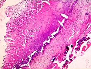
- Updated high quality labeled photomicrographs now which can be used as an Atlas
- Simple, precise and student-friendly text
- Point-wise presentation for easy learning and quick recapitulation during examination
- Line diagrams for basic understanding of the tissue/organ
- Pencil sketches of sections (haematoxylin and eosin) along with salient points of identification, well integrated with text for understanding technical details of structures at the backdrop of theory
- Practical section comprising of enlarged high quality labeled photomicrographs at the end of each chapter with detailed explanation based on students’ expectation to observe
- Clinical correlation of certain important structures
- Self-assessment exercise at the end of theory for revision of the topics studied
From the Publisher
This book has been written in a concise and easily assimilable style to enable rapid understanding of the mechanism and morphology of disease. It has been structured in a question-answer format that incorporates information in numerous flowcharts and tables that are easy to understand, recall and duplicate in the examination. The new edition is based on Robbins and Cotran Pathologic Basis of Disease, 9E.
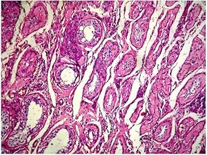
Atrophic testis showing loss of germ cells within the tubules, with peritubular.
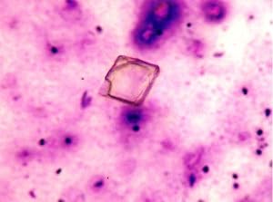
Cholesterol crystal.
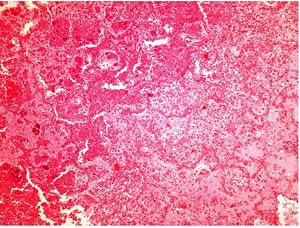
Coagulative necrosis in lung with preservation of alveolar architecture (lung infarct).
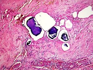
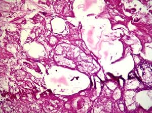
Dystrophic calcification in necrotic foci.
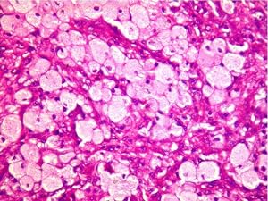
Foamy histiocytes in chronic xanthomatous inflammation.
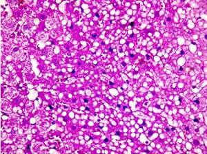
Hepatocytes showing intracellular accumulation of fat appearing as vacuolated to clear cytoplasm and eccentrically displaced nuclei.
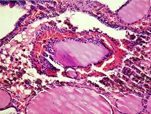
Multinodular goiter showing haemorrhage and haemosiderin deposits.
Photomicrograph of fat necrosis showing foamy histiocytes.
Scab with bacterial colonies (fibrinosuppurative inflammation).
About the Author
An alumnus of Lady Hardinge Medical College, New Delhi, India, Geetika Khanna Bhattacharya is presently working as Professor of Pathology in CIO Laboratory, Vardhman Mahavir Medical College and Safdarjung Hospital, New Delhi, India. In her two decades of teaching experience in prestigious medical colleges in Delhi, she has had a very close association with the medical student fraternity, both as a resident and faculty. She has contributed several articles in national and international journals and has presented several papers/posters in national and international fora. Her current fields of interest include orthopedic pathology and medical education.
Publisher : Elsevier India; 3rd edition (27 July 2016)
Language : English
Paperback : 670 pages
ISBN-10 : 8131244210
ISBN-13 : 978-8131244210
Item Weight : 625 g
Dimensions : 19.61 x 12.53 x 2.01 cm
Country of Origin : India

")
")


 (Canary, 5-Inch by 8-Inch)")

")
")
 -Set of 5 Boxes")
: Supports the National Curriculum, English Exercise Book (Made Easy Workbooks)")


![LSAT Prep Books 2020-2021: Study Guide and 2 LSAT Practice Tests for the LSAC Law School Admission Test [3rd Edition]](https://i1.wp.com/images-na.ssl-images-amazon.com/images/I/71SU+v8dPiL.jpg?w=300&ssl=1 "LSAT Prep Books 2020-2021: Study Guide and 2 LSAT Practice Tests for the LSAC Law School Admission Test [3rd Edition]")